Modulation of Lipid and Glucose Profiles in Hyperglycemic and Hypercholesterolemic Animals by Nanocapsules of Purple Yam Anthocyanin Extract
DOI:
https://doi.org/10.22437/ifstj.v9i2.48120Keywords:
anthocyanin nanocapsules, diabetes mellitus, dyslipidemia, lipid profile, purple yamAbstract
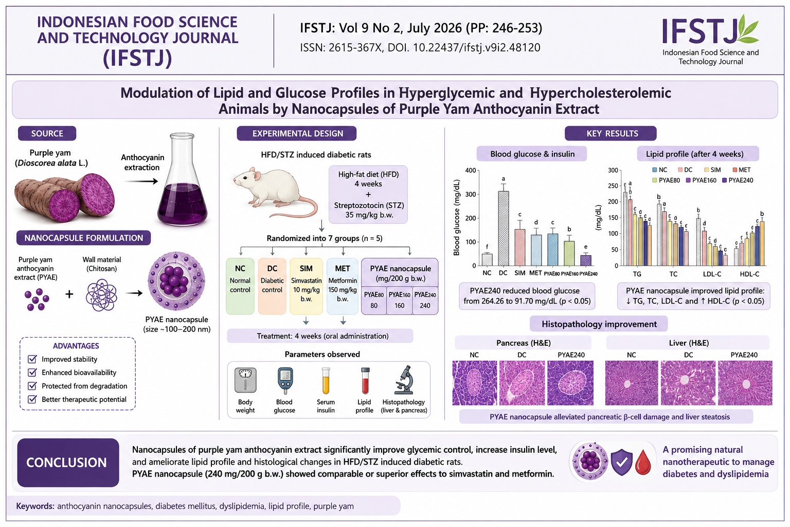
Purple yam, rich in anthocyanins, has potential benefits for dyslipidemia and diabetes. Encapsulation into nanocapsules improves its stability and bioavailability. This study aimed to evaluate the effects of purple yam anthocyanin extract (PYAE) nanocapsules on high-fat diet and streptozotocin (STZ)-induced diabetic rats. Diabetic rats induced with high-fat diet and streptozotocin (STZ) were divided into control and treatment groups receiving simvastatin, metformin, or purple yam anthocyanin extract (PYAE) nanocapsules (80-240 mg/200 g bq) for four weeks. Various parameters were observed, including body weight, blood glucose, serum insulin, lipid profile, and histopathology of the liver and pancreas. The results indicated that treatment with purple yam anthocyanin extract nanocapsules improved body weight, blood glucose levels, insulin levels, and lipid profiles compared to the diabetic rat group. Specifically, the blood glucose level in the group receiving 240 mg/200 g bw decreased significantly from 264.26 mg/dL to 91.70 mg/dL (p<0.05). In addition, body weight and insulin level increased, with insulin rising by 529.89 pg/mL from an initial level of 469.71 pg/mL. Furthermore, PYAE nanocapsules improved the lipid profiles of dyslipidemic rats by reducing triglycerides, cholesterol, and LDL levels while increasing HDL levels (p<0.05). Histopathological examination revealed that PYAE nanocapsule alleviated damage to pancreatic and liver tissues in diabetic rats.References
[1] R. Chakraborty, P. Kalita, and S. Sen, “Phenolic Profile, Antioxidant, Antihyperlipidemic and Cardiac Risk Preventive Effect of Pigmented Black Rice Variety Chakhao poireiton in High-Fat High-Sugar Induced Rats,” Rice Sci., vol. 30, no. 6, pp. 641–651, 2023, doi: 10.1016/j.rsci.2023.08.002.
[2] N. Thongtang, R. Sukmawan, E. J. B. Llanes, and Z. V. Lee, “Dyslipidemia management for primary prevention of cardiovascular events: Best in-clinic practices,” Prev. Med. Reports, vol. 27, no. December 2021, p. 101819, 2022, doi: 10.1016/j.pmedr.2022.101819.
[3] B. Giri, S. Dey, T. Das, M. Sarkar, J. Banerjee, and S. K. Dash, “Chronic hyperglycemia mediated physiological alteration and metabolic distortion leads to organ dysfunction, infection, cancer progression and other pathophysiological consequences: An update on glucose toxicity,” Biomed. Pharmacother., vol. 107, no. July, pp. 306–328, 2018, doi: 10.1016/j.biopha.2018.07.157.
[4] A. Gofur et al., “Evaluation of antioxidant properties from purple tubers and their ability to improve glucose and lipid metabolism in streptozotocin-induced diabetic rats,” Phytomedicine Plus, vol. 4, no. 2, p. 100542, 2024, doi: 10.1016/j.phyplu.2024.100542.
[5] C. J. Packard, J. Boren, and M. R. Taskinen, “Causes and Consequences of Hypertriglyceridemia,” Front. Endocrinol. (Lausanne)., vol. 11, no. May, pp. 1–15, 2020, doi: 10.3389/fendo.2020.00252.
[6] J. Tanoey and H. Becher, “Diabetes prevalence and risk factors of early-onset adult diabetes: results from the Indonesian family life survey,” Glob. Health Action, vol. 14, no. 1, 2021, doi: 10.1080/16549716.2021.2001144.
[7] S. Sapian et al., “The Role of Anthocyanin in Modulating Diabetic Cardiovascular Disease and Its Potential to Be Developed as a Nutraceutical,” Pharmaceuticals, vol. 15, no. 11, 2022, doi: 10.3390/ph15111344.
[8] R. R. Multisona, S. Shirodkar, M. Arnold, and A. Gramza-Michalowska, “Clitoria ternatea Flower and Its Bioactive Compounds: Potential Use as Microencapsulated Ingredient for Functional Foods,” Appl. Sci., vol. 13, no. 4, 2023, doi: 10.3390/app13042134.
[9] K. Shah and P. Shah, “Effect of Anthocyanin Supplementations on Lipid Profile and Inflammatory Markers: A Systematic Review and Meta-Analysis of Randomized Controlled Trials,” Cholesterol, vol. 2018, 2018, doi: 10.1155/2018/8450793.
[10] Z. Fang, D. Wu, D. Yü, X. Ye, D. Liu, and J. Chen, “Phenolic compounds in Chinese purple yam and changes during vacuum frying,” Food Chem., vol. 128, no. 4, pp. 943–948, 2011, doi: 10.1016/j.foodchem.2011.03.123.
[11] J. yu Chen, J. Du, M. li Li, and C. mei Li, “Degradation kinetics and pathways of red raspberry anthocyanins in model and juice systems and their correlation with color and antioxidant changes during storage,” Lwt, vol. 128, no. April, p. 109448, 2020, doi: 10.1016/j.lwt.2020.109448.
[12] J. Chen, X. H. Ma, G. L. Yao, W. T. Zhang, and Y. Zhao, “Microemulsion-based anthocyanin systems: Effect of surfactants, cosurfactants, and its stability,” Int. J. Food Prop., vol. 21, no. 1, pp. 1152–1165, 2018, doi: 10.1080/10942912.2018.1485032.
[13] N. Echegaray et al., “Recent advances in food products fortification with anthocyanins,” Crit. Rev. Food Sci. Nutr., vol. 62, no. 6, pp. 1553–1567, 2022, doi: 10.1080/10408398.2020.1844141.
[14] T. K. O. Rosales and J. P. Fabi, “Nanoencapsulated anthocyanin as a functional ingredient: Technological application and future perspectives,” Colloids Surfaces B Biointerfaces, vol. 218, no. April, 2022, doi: 10.1016/j.colsurfb.2022.112707.
[15] S. Tamaroh and Y. P. Sari, “Antioxidant and Physical Characteristics of Anthocyanin Extract from Purple Yam (Dioscorea alata L.) Nanoencapsulation: Effect of Maltodextrin and Whey Protein Isolate Ratios,” Curr. Res. Nutr. Food Sci., vol. 12, no. 1, pp. 245–253, 2024, doi: 10.12944/CRNFSJ.12.1.20.
[16] A. Murdiati, H. Wuryastuti, Y. Marsono, and E. Harmayani, “Pengaruh Sorbitol-Oleat Polister (SOPE) Terhadap Profil Lipid Serum Tikus Sprague-Dawley,” AGRITECH, vol. 30, no. 1, pp. 18–24, 2010.
[17] R. W, “Analytical reviews in clinical biochemistry: The estimation of urea,” Ann. Clin. Biochem., vol. 29, no. 6, pp. 577–597, 1992, doi: 10.1177/000456329202900301.
[18] M. F. Lopes-Virella, P. Stone, S. Ellis, and J. A. Colwell, “Cholesterol determination in Cholesterol Determination Methods in High-Density Lipoproteins Separated by Three Different,” Clin Chem, vol. 23, no. 5, pp. 882–884, 1997.
[19] Paget GE and B. JM., Toxicity Tests. In: Laurence DR, Bacharach AL, eds. Evaluation of Drug Activities: Pharmacometrics, 1st ed., vol. 1. Academic Press, 1964. doi: https://doi.org/10.1016/B978-1-4832-2845-7.50012-8.
[20] Z. Chen, C. Wang, Y. Pan, X. Gao, and H. Chen, “Hypoglycemic and hypolipidemic effects of anthocyanins extract from black soybean seed coat in high fat diet and streptozotocin-induced diabetic mice,” Food Funct., vol. 9, no. 1, pp. 426–439, 2018, doi: 10.1039/c7fo00983f.
[21] S. Gheibi, K. Kashfi, and A. Ghasemi, “A practical guide for induction of type-2 diabetes in rat: Incorporating a high-fat diet and streptozotocin,” Biomed. Pharmacother., vol. 95, no. 24, pp. 605–613, 2017, doi: 10.1016/j.biopha.2017.08.098.
[22] S. Ziyanok-Demirtas, “Therapeutic potentials of Hibiscus trionum: Antioxidant, anti-lipid peroxidative, hypoglycemic, and hepatoprotective effects in type 1 diabetic rats,” Biomed. Pharmacother., vol. 175, p. 116630, 2024, doi: 10.1016/j.biopha.2024.116630.
[23] A. I. Aladenika YV, Akinjiyan MO, Elekofehinti OO, “Bambusa vulgaris leaf extract inhibits the inflammatory and oxidative pathways in streptozotocin-induced diabetic rats,” J Ethnopharmacol, vol. 339, no. 119116, pp. 1–19, 2025, doi: 10.1016/j.jep.2024.119116.
[24] A. O. Olatunji et al., “Co-administration of Hibiscus sabdariffa linn and Daflon-500® mitigates weight loss, hyperglycemia, hematological and oxidative changes in male rats with type-2 diabetes,” Phytomedicine Plus, vol. 5, no. 1, p. 100699, 2025, doi: 10.1016/j.phyplu.2024.100699.
[25] H. J. Wang et al., “Low dose streptozotocin (STZ) combined with high energy intake can effectively induce type 2 diabetes through altering the related gene expression,” Asia Pac. J. Clin. Nutr., vol. 16, no. SUPPL.1, pp. 412–417, 2007.
[26] D. D. Herrera-Balandrano et al., “Hypoglycemic and hypolipidemic effects of blueberry anthocyanins by AMPK activation: In vitro and in vivo studies,” Redox Biol., vol. 46, p. 102100, 2021, doi: 10.1016/j.redox.2021.102100.
[27] Y. Liu et al., “Anthocyanins’ effects on diabetes mellitus and islet transplantation,” Crit. Rev. Food Sci. Nutr., vol. 63, no. 33, pp. 12102–12125, 2023, doi: 10.1080/10408398.2022.2098464.
[28] B. Y. Sharma S, Pandita G, “Anthocyanin: Potential tool for diabetes management and different delivery aspects,” Trends Food Sci Technol, vol. 140, no. 104170, pp. 1–17, 2023, doi: 10.1016/j.tifs.2023.104170.
[29] R. P. Munshi, S. G. Joshi, and B. N. Rane, “Development of an experimental diet model in rats to study hyperlipidemia and insulin resistance, markers for coronary heart disease,” Indian J. Pharmacol., vol. 46, no. 3, pp. 270–276, 2014, doi: 10.4103/0253-7613.132156.
[30] J. Soppert, M. Lehrke, N. Marx, J. Jankowski, and H. Noels, “Lipoproteins and lipids in cardiovascular disease: from mechanistic insights to therapeutic targeting,” Adv. Drug Deliv. Rev., vol. 159, pp. 4–33, 2020, doi: 10.1016/j.addr.2020.07.019.
[31] H. Song, X. Shen, Y. Zhou, and X. Zheng, “Black rice anthocyanins alleviate hyperlipidemia, liver steatosis and insulin resistance by regulating lipid metabolism and gut microbiota in obese mice,” Food Funct., vol. 12, no. 20, pp. 10160–10170, 2021, doi: 10.1039/d1fo01394g.
[32] T. C. Peixoto et al., “Cranberry (Vaccinium macrocarpon) extract treatment improves triglyceridemia, liver cholesterol, liver steatosis, oxidative damage and corticosteronemia in rats rendered obese by high fat diet,” Eur. J. Nutr., vol. 57, no. 5, pp. 1829–1844, 2018, doi: 10.1007/s00394-017-1467-2.
[33] C. Garcia and C. N. Blesso, “Antioxidant properties of anthocyanins and their mechanism of action in atherosclerosis,” Free Radic. Biol. Med., vol. 172, no. May, pp. 152–166, 2021, doi: 10.1016/j.freeradbiomed.2021.05.040.
[34] K. Thompson, W. Pederick, and A. B. Santhakumar, “Anthocyanins in obesity-associated thrombogenesis: A review of the potential mechanism of action,” Food Funct., vol. 7, no. 5, pp. 2169–2178, 2016, doi: 10.1039/c6fo00154h.
[35] D. Li, Y. Zhang, Y. Liu, R. Sun, and M. Xia, “Purified anthocyanin supplementation reduces dyslipidemia, enhances antioxidant capacity, and prevents insulin resistance in diabetic patients,” J. Nutr., vol. 145, no. 4, pp. 742–748, 2015, doi: 10.3945/jn.114.205674.
[36] K. S. Stote et al., “Effect of blueberry consumption on cardiometabolic health parameters in men with type 2 diabetes: An 8-week, double-blind, randomized, placebo-controlled trial,” Curr. Dev. Nutr., vol. 4, no. 4, pp. 1–10, 2020, doi: 10.1093/CDN/NZAA030.
[37] A. Aboonabi, R. R. Meyer, A. Gaiz, and I. Singh, “Anthocyanins in berries exhibited anti-atherogenicity and antiplatelet activities in a metabolic syndrome population,” Nutr. Res., vol. 76, pp. 82–93, 2020, doi: 10.1016/j.nutres.2020.02.011.
[38] C. Liu, J. Sun, Y. Lu, and Y. Bo, “Effects of anthocyanin on serum lipids in dyslipidemia patients: A systematic review and meta-analysis,” PLoS One, vol. 11, no. 9, pp. 1–11, 2016, doi: 10.1371/journal.pone.0162089.
[39] Y. P. Hwang et al., “Purple sweet potato anthocyanins attenuate hepatic lipid accumulation through activating adenosine monophosphate-activated protein kinase in human HepG2 cells and obese mice,” Nutr. Res., vol. 31, no. 12, pp. 896–906, 2011, doi: 10.1016/j.nutres.2011.09.026.
[40] H. Aboufarrag, W. J. Hollands, J. Percival, M. Philo, G. M. Savva, and P. A. Kroon, “No Effect of Isolated Anthocyanins from Bilberry Fruit and Black Rice on LDL Cholesterol or other Biomarkers of Cardiovascular Disease in Adults with Elevated Cholesterol: A Randomized, Placebo-Controlled, Cross-Over Trial,” Mol. Nutr. Food Res., vol. 66, no. 21, pp. 1–9, 2022, doi: 10.1002/mnfr.202101157.
[41] P. Prangthip et al., “Amelioration of hyperglycemia, hyperlipidemia, oxidative stress and inflammation in steptozotocin-induced diabetic rats fed a high fat diet by riceberry supplement,” J. Funct. Foods, vol. 5, no. 1, pp. 195–203, 2013, doi: 10.1016/j.jff.2012.10.005.
[42] Y. M. Yang, Y. E. Cho, and S. Hwang, “Crosstalk between Oxidative Stress and Inflammatory Liver Injury in the Pathogenesis of Alcoholic Liver Disease,” Int. J. Mol. Sci., vol. 23, no. 2, 2022, doi: 10.3390/ijms23020774.
Downloads
Published
Issue
Section
License
Copyright (c) 2026 Indonesian Food Science and Technology Journal

This work is licensed under a Creative Commons Attribution 4.0 International License.